

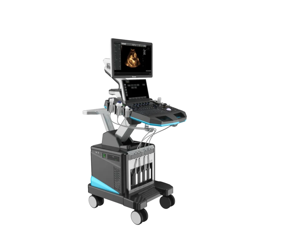
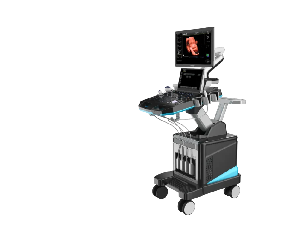
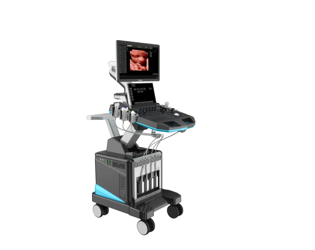
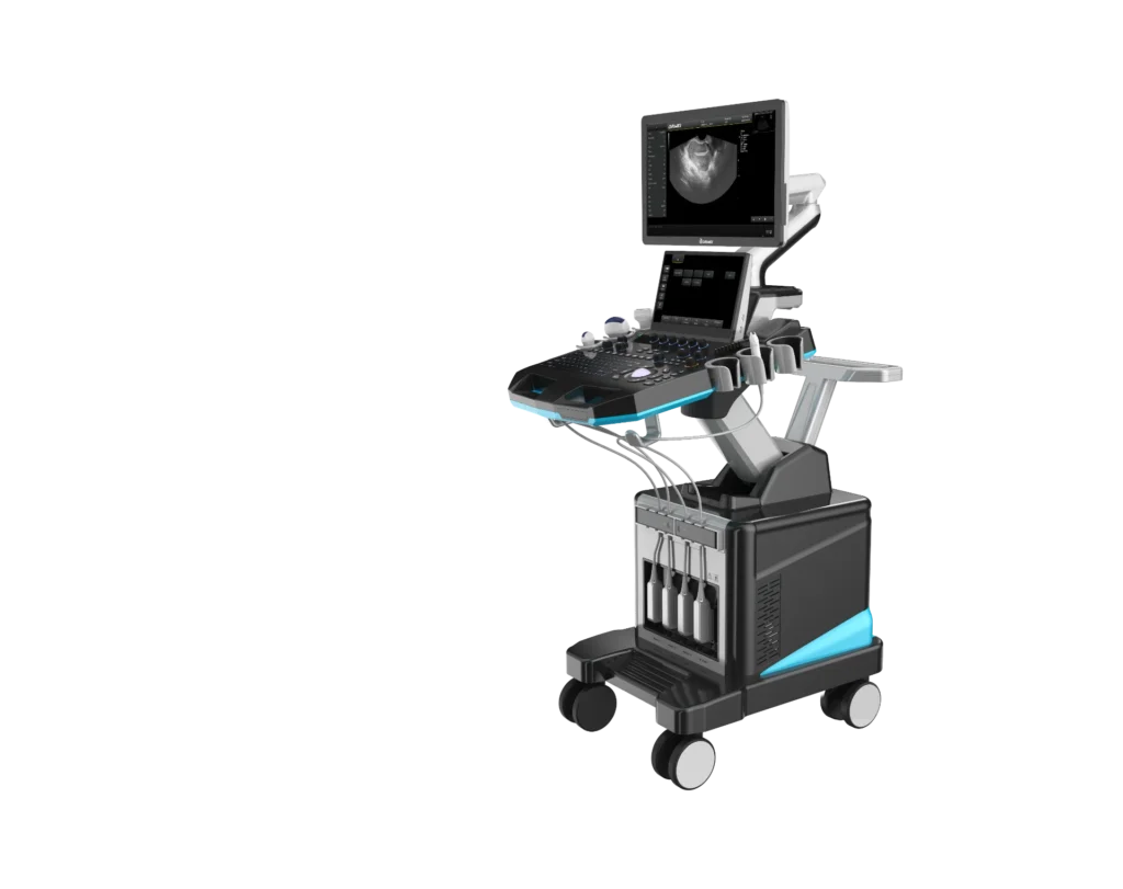
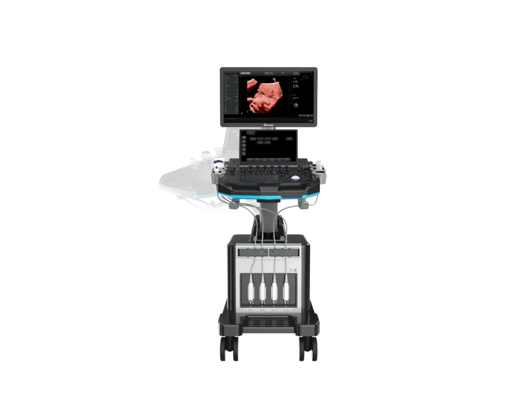
SM-T30 Trolley Color Ultrasonic Diagnostic Apparatus
1. Advanced 3D/4D D-Live & Smartface Technology
Break through the limits of traditional grayscale ultrasound. Our 3D/4D D-Live technology delivers more intuitive, three-dimensional and hyper-realistic imaging. It performs excellently in fetal abnormality screening, with wider observation range and ultra-clear picture quality.
Silhouette Function
Generate unique transparent 3D fetal anatomical images to streamline primary anatomical assessment. Different from conventional ultrasound, this module visualizes the complete internal anatomical structure of the fetus for more comprehensive evaluation.
Volumetric 4D Probe Support
Equipped with dedicated volumetric probe to unlock full 3D/4D imaging capacity for obstetric clinical use.
2. Wide Clinical Applications
Suitable for multi-department routine diagnosis:
Abdominal
Obstetrics
Pediatrics
Musculoskeletal
Gynecology
Small organs
Vessels
Kidney
Cardiac
3. Ergonomic Flexible Height & Angle Adjustment
Designed under strict ergonomic standards. The operation panel supports 18cm vertical lifting, 110° horizontal rotation, and multi-angle tilting, allowing doctors to customize the most comfortable operating posture, reducing fatigue and improving diagnostic efficiency.
4. Humanized Operation Panel Design
21.5-inch medical high-definition main display
13.3-inch large-size touch screen, flip angle adjustable from 0° to 45°
Operation panel: 110° horizontal rotation + 18cm vertical lifting
Detachable probe hanging slots for tidy storage
4 active probe interfaces to connect multiple transducers at once
Flexible silent casters with directional locking function for easy ward movement
5. Professional Specialized Measurement Packages
Built-in dedicated measurement software packages for Abdomen, Obstetrics, Urology, Gynecology and more departments. Provide clinicians with standardized, quantitative data to assist scientific and accurate diagnosis.
6. High-Speed Platform to Accelerate Workflow
Advanced YH+ Operating Platform
8GB high-speed RAM + 512GB solid-state SSD
Powerful real-time data processing capacity
Smooth human-machine interaction and uninterrupted scanning
Sub-array Beamforming Technology
Deliver ultra-high resolution images with exquisite tissue details, significantly lifting the accuracy of clinical diagnosis.
7. Complete Hospital Data Sharing Functions
Real-time LAN Sharing
Transmit ultrasound videos and images to hospital PCs instantly via local area network.
DICOM 3.0 Standard Compatible
Seamlessly connect with hospital PACS system for fast image transmission and file archiving.
Optimized Image Presets
Pre-set optimal imaging parameters for different examinations to avoid repeated adjustments and save scanning time.
One-Click Image Optimization
Instantly optimize imaging quality with one tap to shorten operating procedures.
Automatic Spectrum Envelope Analysis
Auto-calculate core hemodynamic parameters: PSV, EDV, RI, PI, S/D, ACC, HR etc., output complete diagnostic data automatically.
8. Premium Imaging Technologies for Clearer Visualization
Trapezoidal Imaging
Extended trapezoidal field of view based on standard rectangular scanning frame, expanding left and right detection range to cover larger tissue areas.
Directional Power Doppler Imaging (DPDI)
Adopt high-resolution tiny sampling volume to generate dual-color blood flow directional signals. Reduce color blooming artifact, accurately reflect lesion size and make up for ordinary power Doppler’s deficiency of no flow direction display.
Tissue Harmonic Imaging (THI)
Boost tissue contrast and spatial resolution, eliminate near-field artifacts. Ideal for cardiovascular and abdominal diagnosis, accurately defining lesion boundary for hard-to-image patients.
Micron Imaging
Real-time edge signal tracking of different tissues to enhance contour definition. Synchronously optimize intra-tissue pixel signals, perfectly fuse edge and internal information, restore delicate 2D images with superior contrast.
Intelligent Clean Filter
Filter and extract valid signals across all frequency bands and tissue depths. Perform targeted signal correction and noise suppression to output high-fidelity, low-noise ultrasound images.
9. Optional Matching Probes for Full Clinical Coverage
Probe Type Main Application Scenarios
Convex Probe Abdomen, Obstetrics, Gynecology
Linear Probe Vascular, Musculoskeletal System
Phased Array Probe Cardiac chambers, cardiac function, pericardial effusion
4D Volume Probe Fetal 3D/4D obstetric scanning
Trans-vaginal Probe Obstetrics, Gynecology, Urology
Trans-rectal Probe Prostate examination
Micro-convex Probe Small organs, pediatric organs