
Feeling anxious about breast cancer screening[^1]? The guidelines can be confusing, leaving you unsure about the next steps. We're here to provide clear, simple answers for your peace of mind.
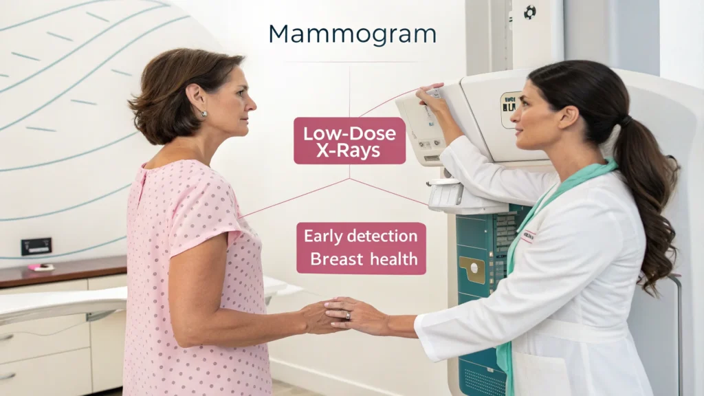
A mammogram[^2] is a low-dose X-ray of the breast used to detect early signs of cancer. Getting one is a key part of preventive health for women, especially after age 40. It's the most effective tool we have for finding breast cancer early.

This guide will walk you through everything you need to know. We'll cover the types of mammogram[^2]s, when to get one, and what to expect. My goal as a medical equipment professional is to demystify the process, so you feel confident and informed. Let's start with the basics.
What Is a Mammogram, Really?
You've heard the term "mammogram[^2]," but what does it actually involve? It sounds intimidating, making you hesitant to schedule one. Let's break down this simple, life-saving procedure.
A mammogram[^2] is a specialized medical imaging test that uses low-dose X-rays to examine breast tissue. Its primary purpose is to aid in the early detection[^3] and diagnosis of breast diseases, especially cancer, before a lump can even be felt.

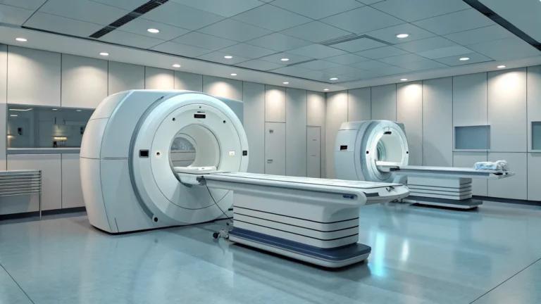
From my perspective in the medical supply industry, I see these machines as more than just hardware. They are the frontline defense in women's health. The technology is designed for one specific purpose: to find cancer when it is most treatable.
How the Technology Works
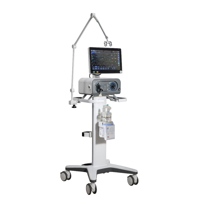
A mammogram[^2] machine has several key parts. An X-ray tube generates a low dose of radiation. The breast is briefly compressed between two plates, called paddles. This compression is crucial because it spreads out the breast tissue, allowing for a clearer image with less radiation. A digital detector then captures the image, which is sent to a radiologist[^4] for review. This entire process is designed to find tiny abnormalities, like microcalcifications or small masses, that could be the earliest signs of cancer.
The Goal: Early Detection
There are two main reasons to get a mammogram[^2].
| Type of Mammogram | Purpose | Who Is It For? |
|---|---|---|
| Screening | To check for signs of cancer in women with no symptoms. | Routine check-ups, usually done annually or biennially. |
| Diagnostic | To investigate a specific problem, like a new lump or an abnormal screening result. | When there is a specific concern that needs a closer look. |
As a provider, we ensure hospitals have access to both types of systems to build a complete breast health program[^5].
What Are the Main Types of Mammograms Today?
Is a 3D mammogram[^6]m](https://en.wikipedia.org/wiki/Mammography)[^2] better than a 2D one? The options can be overwhelming, causing confusion about which test is right for you. We'll clarify the differences simply.
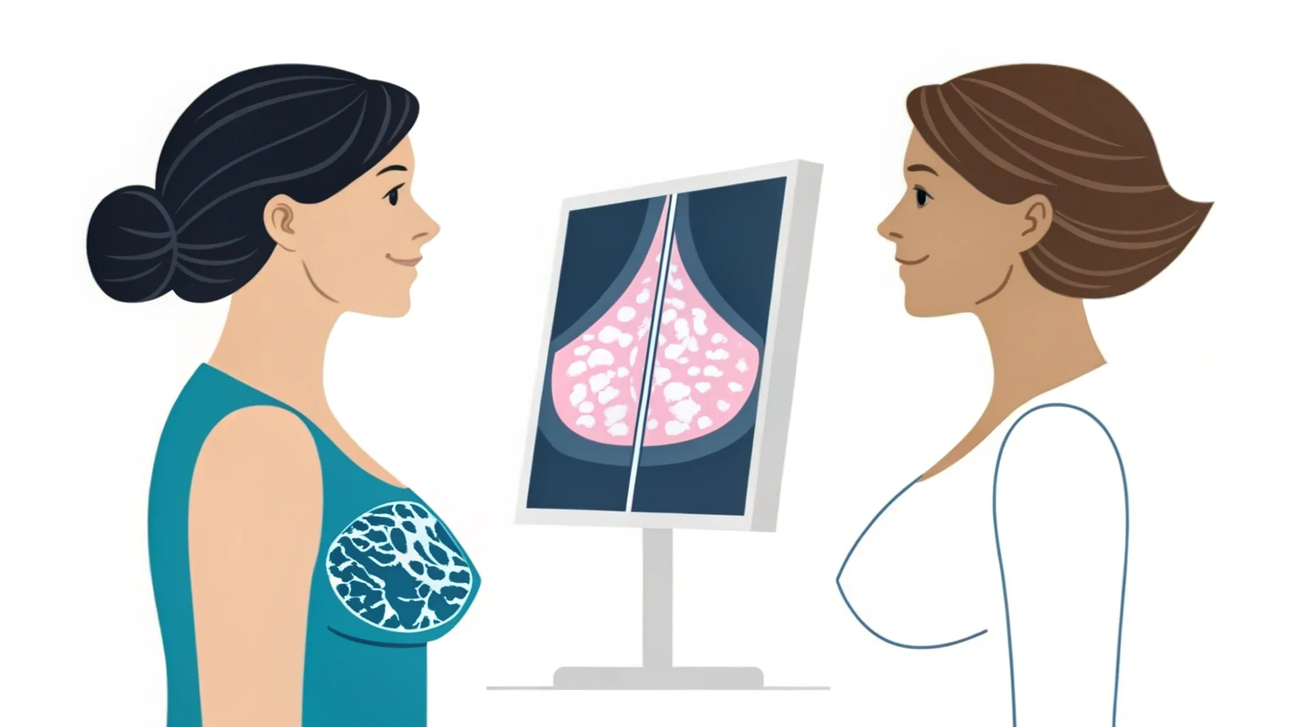
The two main types are 2D (conventional) and 3D (tomosynthesis) mammogram[^2]s. 3D mammogram[^6]m](https://en.wikipedia.org/wiki/Mammography)[^2]s take multiple images to create a clearer, more detailed picture of the breast, which is especially helpful for women with dense breast tissue.

In our business, we've seen a major shift toward 3D mammography. Hospitals and clinics are upgrading because the clinical benefits are becoming very clear. It helps radiologist[^4]s do their job better, which is good for everyone.
2D vs. 3D: A Closer Look
The standard for many years was the 2D mammogram[^7]m](https://en.wikipedia.org/wiki/Mammography)[^2], which takes two X-rays of each breast from different angles. It's effective, but it produces a flat image. This can sometimes be a problem in women with dense breasts, where overlapping tissue can hide a small cancer or create a shadow that looks like one. 3D mammography, or tomosynthesis, addresses this. It takes many low-dose X-rays as the machine moves in an arc over the breast. A computer then reconstructs these images into a 3D-like view, allowing the radiologist[^4] to examine the tissue one thin layer at a time.
| Feature | 2D Mammography | 3D Mammography (Tomosynthesis) |
|---|---|---|
| Image | A single, flat image. | Multiple thin "slices" of the breast. |
| Detection Rate | Good | Better, especially in dense breasts. |
| False Positives | Higher | Lower (fewer "call-backs"). |
| Patient Time | Slightly faster. | A few seconds longer per view. |
Advanced Screening Options
For some specific cases, even more advanced technology is available. Contrast-Enhanced Mammography (CEM) involves an injection of iodine-based contrast dye before the mammogram[^2]. Cancerous tumors often have more blood vessels, so they "light up" on the image after absorbing the dye. This is not a routine screening tool but can be very helpful for solving difficult diagnostic questions.
Who Should Get a Mammogram—and When?
Screening guidelines seem to change constantly. This uncertainty makes it hard to know if you need a mammogram[^2] now or later. Let's review the current expert recommendations.
Generally, women with an average risk of breast cancer should start getting annual mammogram[^2]s at age 40. However, your personal and family medical history may require an earlier or different screening schedule. Always discuss your specific situation with your doctor.
The debate over screening guidelines[^8] can be confusing. My advice is always to focus on your personal situation. General guidelines are a starting point, not a final rule. A conversation with your healthcare provider is the most important step.
General Screening Guidelines
Different health organizations have slightly different recommendations, but the overall message is similar. Early and regular screening saves lives. Here is a simplified overview for women at average risk.
| Age Group | Common Recommendation |
|---|---|
| Under 40 | Screening not usually recommended unless at high risk. |
| 40–49 | Discuss with your doctor. Most U.S. guidelines now advise starting annual screening at 40. |
| 50–74 | Annual or biennial (every two years) mammogram[^2]s are recommended. |
| 75+ | Continue screening as long as you are in good health and would seek treatment if cancer were found. |
Risk Factors to Consider
You may be at a higher-than-average risk if you have certain factors. These include a personal history of breast cancer, a strong family history (especially in a mother or sister), or a known genetic mutation like BRCA1 or BRCA2. Other factors are having received radiation therapy to the chest before age 30 or having very dense breasts. If any of these apply to you, your doctor may recommend starting mammogram[^2]s earlier, getting them more often, or adding other tests like a breast MRI[^9] or ultrasound.
How Can You Best Prepare for Your Mammogram?
Worried about your mammogram[^2] appointment? Not knowing how to prepare can add to the stress of the test itself. Follow these simple steps for a smoother experience.
On the day of your mammogram[^2], do not wear deodorant, antiperspirant, or lotion under your arms or on your breasts. These products can appear as white spots on the X-ray. Wear a two-piece outfit for convenience, as you'll need to undress from the waist up.

Having worked with many imaging centers, I know that good preparation makes a big difference. It helps the technologist get the best possible images and makes the entire process faster and more comfortable for you.
Before Your Appointment
A little planning goes a long way. If you have a choice, try to schedule your mammogram[^2] for the week after your menstrual period. Your breasts are usually least tender at this time. When you make the appointment, tell the scheduler if you have breast implants or any physical limitations that might make positioning difficult. If you have had mammogram[^2]s at another facility before, arrange to have those images sent to the new facility. The radiologist[^4] will want to compare your new mammogram[^2] to your old ones.
On the Day of Your Test
Your main job is to avoid certain products.
| Preparation Step | Why It's Important |
|---|---|
| Do not use deodorant/lotion | Metallic particles in these products can show up on the X-ray and look like calcifications. |
| Wear a two-piece outfit | You will only need to remove your top and bra, which is more comfortable and convenient. |
| Inform the technologist | Tell them about any new lumps, skin changes, or areas of concern you have noticed. |
What to Bring
Bring your insurance card and any required paperwork. It's also helpful to have the name and address of your primary care doctor so the results can be sent to them. Most importantly, bring any questions you have.
What Actually Happens During the Test?
The thought of the mammogram[^2] procedure itself can be daunting. Fear of pain or discomfort might even make you delay it. Let's walk through the process step-by-step.
During the test, a technologist will position one of your breasts on a platform. A clear plastic plate will press down to compress the breast for a few seconds while the X-ray is taken. This is done to spread the tissue and get a clear picture.

I've spoken with many technologists who perform these exams every day. They understand that patients can be nervous. Their main goal is to make the experience as quick and comfortable as possible while getting high-quality images. Don't be afraid to speak up if you are in pain.
Step 1: Changing and Preparation
You will be given a gown and asked to undress from the waist up. The technologist will confirm your medical history and ask if you have any current breast problems. This is your chance to point out any lumps, tender spots, or skin changes.
Step 2: Positioning
You and the technologist will be the only ones in the room during the mammogram[^2]. The technologist, who is specially trained in mammography, will place one of your breasts on the machine's imaging platform. They will adjust your body and head to get the correct view.
Step 3: Compression and Imaging
The technologist will then lower a clear plastic plate to compress your breast. This compression is necessary for a few reasons: it evens out the breast thickness for a clearer picture, spreads the tissue so small abnormalities are not hidden, and holds the breast still to prevent a blurry image. The compression only lasts for a few seconds while the X-ray is taken. Most women find it uncomfortable, but few describe it as truly painful. The process is then repeated for the other breast. A routine screening usually involves two views of each breast.
The Role of Artificial Intelligence (AI)
AI is the most immediate change coming to mammography. AI programs are being trained on millions of mammograms to recognize patterns that are suspicious for cancer. The goal is not to replace radiologists but to assist them. An AI can act as a "second reader," flagging areas of concern that a human might miss. This can improve accuracy and speed up the reading process, which is critical given the shortage of radiologists.
New Imaging Techniques
Beyond AI, new types of imaging are being developed. We already discussed Contrast-Enhanced Mammography (CEM). Another is Molecular Breast Imaging (MBI), a nuclear medicine technique that is very good at finding cancers in dense breasts. These are currently used as secondary tools, but they may become more common in the future for specific patient groups.
Beyond Imaging: Liquid Biopsies
The ultimate goal is to find cancer even earlier, perhaps without any imaging at all. "Liquid biopsies" are blood tests that can detect tiny fragments of DNA shed by cancer cells. While not yet ready for use as a general screening tool, research is advancing rapidly. One day, a simple blood test could tell you if you are at high risk or even have an early, hidden cancer, which would then trigger a more focused imaging test.
What Questions Should You Ask Your Doctor?
Walking into your doctor's office can be overwhelming. You might forget what to ask, leaving you with unanswered questions. Being prepared helps you advocate for your health.
Ask your doctor when you should start mammograms based on your personal risk. Also ask if you have dense breasts and if you need any additional screening, like an ultrasound. Being an active participant in your healthcare is key.

Empowering patients with information is just as important as providing doctors with good equipment. An informed patient is a partner in their own health. Never feel shy about asking questions until you understand the answers.
Before Your First Mammogram
- Based on my family history and personal health, what is my risk for breast cancer?
- At what age should I start getting mammograms?
- How often should I be screened?
- Is there anything I should do to prepare for my first test?
During Your Regular Check-ups
- My last mammogram said I have dense breasts. What does that mean for me?
- Do I need any extra screening tests, like a breast ultrasound or MRI?
- Are there any lifestyle changes I can make to lower my risk?
After an Abnormal Result
- What does my BI-RADS score mean?
- What is the next step? What additional tests do I need?
- How soon should I have these tests done?
- What is the likelihood that this is actually cancer?
Being prepared with these questions will help you have a productive conversation and ensure you are an active manager of your own health journey.
Conclusion
Regular mammograms are a vital tool for early breast cancer detection. Understanding the process and discussing it with your doctor empowers you to take control of your health.
[^1]: Stay informed about breast cancer screening guidelines to ensure timely check-ups and peace of mind. [^2]: Understanding mammograms is crucial for early cancer detection; explore this link for detailed insights. [^3]: Learn why early detection can save lives and how it impacts treatment options. [^4]: Learn about the role of radiologists in interpreting mammograms and ensuring accurate results. [^5]: Discover the components of a complete breast health program for proactive care. [^6]: Explore the advantages of 3D mammograms for better detection, especially in dense breast tissue. [^7]: Discover the differences between 2D and 3D mammograms to make informed health decisions.